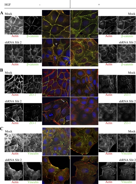
Figure 7.
Distribution of adhesion markers in MDCK cells with normal or reduced levels of Slit-2. Double staining for actin and molecular markers of cell–cell contacts (A and B) or cell-substrate adhesion (C) in mock and shRNA-SLIT-2 cells in the presence (+) or absence (−) of HGF (40 ng/ml for 60 min). The lateral enrichment of β-catenin (A) and ZO-1 (B) is basally reduced in Slit-2–deficient cells (arrowhead for β-catenin and arrows for ZO-1) and further decreases after short-term HGF stimulation. Similarly, vinculin immunoreactivity (C) at focal contacts is impaired in Slit-2–deficient cells, with further cytoplasmic redistribution after HGF. β-Catenin and ZO-1 staining is from apical optical sections; vinculin staining is from basal sections. Bar, 4 μm (color) and 6 μm (black and white).

