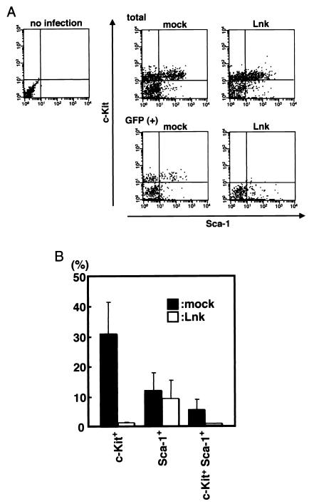
FIG. 3.
Lnk-driven inhibition of the appearance of c-Kit on cultured AGM cells. (A) E11.5 AGM cells were cultured with SCF, bFGF, and OSM. On day 2 of culture, cells were infected with GFP or Lnk retrovirus. On day 7 of culture, Lin− cells were separated from nonadherent cells with magnetic microbeads. Purified cells were stained with anti-c-Kit and Sca-1 antibodies and analyzed by flow cytometry. In each flow cytometric profile, 3 × 10³ (total cells, upper three panels) or 5 × 10² (GFP+ cells, lower two panels) events are recorded. (B) The percentage of c-Kit+ and Sca-1+ cells in the 5 × 10² GFP+ cells was determined. Error bars indicate the standard error of the mean (n = 5).

