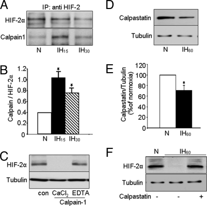
Fig. 4.
Evidence for interactions of HIF-2α with calpain 1. (A) PC12 cells were exposed to normoxia or IH15 and IH30. Immunocomplexes precipitated with anti-HIF-2α were analyzed on 6% SDS/PAGE gels and probed with anti-HIF-2α (Top) and anti-calpain-1 (Bottom) antibodies (n = 3). (B) Densitometric analysis of the immunoblots from 3 experiments expressed as ratio of calpain to HIF-2α. (C) HIF-2α protein was analyzed by immunoblot in PC12 cell lysates incubated for 15 min with purified calpain-1 (3 mg/mL) in the presence of 1 mM CaCl2 or 1 mM CaCl2 plus 2 mM EDTA. (n = 3). (D and E) Immunoblot and densitometric data showing decreased calpastatin protein expression in PC12 cells exposed to IH60 (n = 5) compared with cells exposed to normoxia (N). (F) Representative example of an immunoblot showing that 2 μM calpastatin rescues IH60-evoked HIF-2α degradation (n = 3). Tubulin expression was determined as control for protein loading in C, D, and F.

