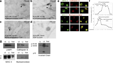
Figure 3.
EDBs are myeloperoxidase positive and TGF-α negative. A–D) Ultrathin cryosections of monocytes and immunogold labeling: HLA-DR (10 nm gold) and myeloperoxidase (5 nm gold) (A–C); HLA-DR (10 nm gold) and TGF-α (5 nm gold) (D). E) Confocal immunostaining for MHC class II and cathepsin G (top panels) and myeloperoxidase (bottom panels). F) β-Hexosaminidase activity measured in each fraction (1 ml) of a 10/27% 2-step Percoll gradient to separate lysosomes and late endosomes. G) Western blot analysis of pulled fractions 6–7 of the 27% Percoll gradient (lysosomes, Ly), fractions 3–4 of the 10% Percoll gradient (late endosomes, LE) and total cell lysates (total). Membranes were probed using a rabbit serum recognizing both α and β subunits of HLA-DR1 or mAb specific for Lamp-1, cathepsin S, invariant chain, and myeloperoxidase. Monocyte preparations from 4 different subjects were analyzed.

