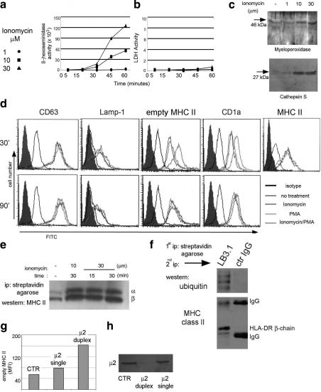
Figure 4.
EDBs behave as secretory lysosomes. A) β-Hexosaminidase detection in the cell culture supernatant after treatment of monocytes with different concentrations of ionomycin for different times. B) Lactic-dehydrogenase detection in the cell culture supernatant after treatment of monocytes with different concentrations of ionomycin for different times. C) Western blot analysis for cathepsin S and myeloperoxidase secreted in the culture supernatant after ionomycin treatment of primary monocytes. D) Surface immunostaining of monocytes untreated or after ionomycin, phorbol-12-myristate-13-acetate (PMA), or ionomycin/PMA treatment. E) Western blot analysis of MHC class II proteins following ionomycin treatment, surface biotinilation, and streptavidin agarose pulldown. F) Western blot analysis of ubiquitinated surface biotinylated MHC class II proteins. G) Surface MHC class II protein staining of monocytes nontransfected (control) or transfected with single or duplex siRNA to the AP-2 adaptor complex. Data are reported as mean fluorescence intensity. H) Western blot analysis of monocytes nontransfected (control) or transfected with single or duplex siRNA. Membrane is probed with the μ2 mAb. Each reported experiment was performed between 3 and 5 times.

