Figure 2.
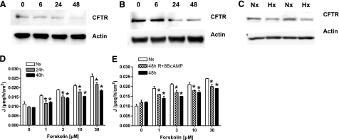
Epithelial hypoxia attenuates CFTR protein and function. A, B) Caco-2 (A) and T84 cells (B) were exposed to normoxia or indicated times of hypoxia, and cell lysates were examined by Western blot using specified antibodies. Blots are representative of 3 independent experiments. C) In a whole-body murine hypoxia model (Hx; 4 h), Western blot analysis for CFTR was performed on small intestinal mucosal scrapings. β-Actin served as control. Western blots are representative for 3 separate experiments; n = 2/condition. Iodine efflux in hypoxic epithelia was measured colorimetrically. D) T84 cells were exposed to indicated times of hypoxia, and efflux of loaded iodine was measured. E) T84 cells were either exposed to normoxia (empty bars) and hypoxia (48 h; filled bars) alone or exposed to hypoxia and reoxygenated in presence of 8-bromo-cAMP before measuring iodine efflux (48h+R+cAMP; checkered bars). Data are expressed as coefficient of iodine secretion J ± sem; *P ≤ 0.05 vs. normoxia.

