Figure 2.
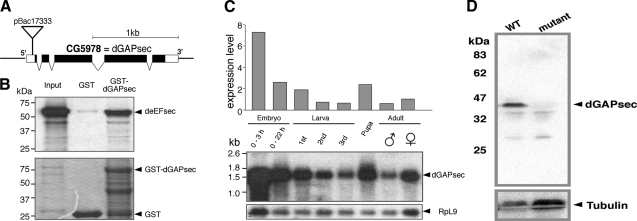
Identification of the Drosophila dGAPsec gene and lack of protein in the mutant. A) Genomic organization of dGAPsec (CG5978) as established by sequence comparison of genomic DNA (http://flybase.bio.indiana.edu/) and cDNA isolates. pBac17333, position of the pBac insertion; black bars, coding sequences; open bars, untranslated regions. The processed CG5978 transcript is 1.541 kb in length. B) Full-length GST-dGAPsec fusion protein interacts with deEFsec. GST-pulldown with in vitro translated 35S-labeled deEFsec (input) and bacterially produced GST (GST) as well as GST-dGAPsec (GST-dGAPsec). Top panel: autoradiograph of 35S-labeled protein; bottom panel: Coomassie brilliant blue-stained gel. C) Developmental profile of dGAPsec transcripts. The Northern blot was initially probed for deEFsec mRNA (17), stripped, and reprobed with a dGAPsec probe. The quantified profile is shown above the Northern blot (middle panel). Bottom panel: RpL9 transcripts served as reference for mRNA quantification. D) Anti-dGAPsec antibody staining of Western blots (WT, wild type; mutant, homozygous dGAPsec mutant flies) shows that the protein is absent in mutants. Protein loading of blots was controlled by anti-tubulin antibody staining.

