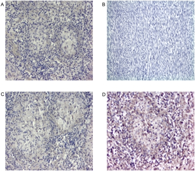
Figure 1. Positive and negative control staining in human tissue.
Paraffin-embedded tissue was stained using ISH for mRNA expression. Negative and sense probe controls were repeated each time lung tissue was stained for cytokine expression. In the first control (A) lung granulomatous tissue was not stained with probe. The absence of brown staining proves that there was no nonspecific staining. Magnification 200×. In the second control (B) anti-sense probe was applied to tissue known not to produce cytokines (sarcoma). The absence of brown staining proves that there was no nonspecific staining. Magnification 400×. In the third control (C) granulomatous lung tissue was stained with sense probe. The absence of brown staining represents specificity. Magnification 200×. In the last control (D) granulomatous lung tissue was stained with the anti-sense β-actin probe. Brown staining indicates the presence of non-degraded mRNA. β-actin expression is diffusely positive. Magnification 200×.

