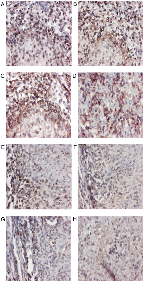
Figure 4. Cytokine mRNA expression in HIV positive and negative pleural tuberculous granulomas.
Paraffin-embedded tissue was stained using ISH for mRNA expression. Representative sections of pleural needle biopsies from HIV positive (A–D) patients are shown. (A) Staining with the anti-sense IL-12 probe shows that over 75% of cells are positive for IL-12 mRNA expression. Magnification 400×. (B) Staining with the anti-sense IFN-γ probe shows that over 75% of cells are positive for IFN-γ mRNA expression. Magnification 400×. (C) Staining with the anti-sense TNF-α probe shows that over 75% of cells are positive for TNF-α mRNA expression. Magnification 400×. (D) Staining with the anti-sense IL-4 probe shows that over 75% of cells are positive for IL-4 mRNA expression. Magnification 400×. Representative sections of pleural needle biopsies from HIV negative (E–H) patients are shown. (E) Staining with the anti-sense IL-12 probe shows that 25%–75% of cells are positive for IL-12 mRNA expression. Magnification 100×. (F) Staining with the anti-sense IFN-γ probe shows that 25%–75% of cells are positive for IFN-γ mRNA expression. Magnification 100×. (G) Staining with the anti-sense TNF-α probe shows that 25%–75% of cells are positive for TNF-α mRNA expression. Magnification 100×. (H) Staining with the anti-sense IL-4 probe shows that less than 25% of cells are positive for IL-4 mRNA expression. Magnification 100×.

