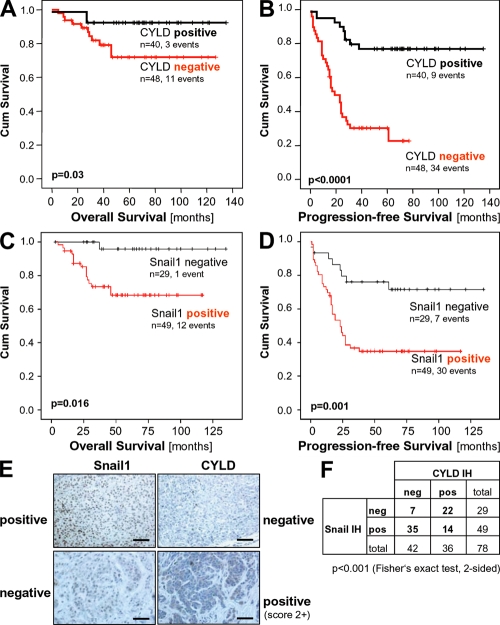
Figure 7.
CYLD expression in melanoma has prognostic implication. Kaplan-Meier curves for overall survival (A) and progression-free survival (B) in melanoma patients with a positive immunosignal for CYLD (CYLD positive) or without detectable CYLD protein expression (CYLD negative). CYLD immunohistochemical staining of primary malignant melanoma tissue was performed using a tissue microarray consisting of 88 cases. Investigation of CYLD protein expression was informative in all specimens. Kaplan-Meier curves for overall survival (C) and progression-free survival (D) in melanoma patients with a positive immunosignal for Snail1 (Snail1 positive) or without detectable Snail1 protein expression (Snail1 negative). Snail1 immunohistochemical staining of primary malignant melanoma tissue was performed using a tissue microarray consisting of 88 cases. Immunohistochemical analysis of Snail1 was informative in 78 samples. (E) Examples of tissues with positive and negative CYLD and Snail1 staining, respectively. Bar, 100 μm. (F) Comparison of CYLD and Snail1 immunoreactivity. An inverse correlation of CYLD and Snail1 staining was observed.

