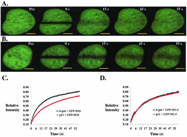
FIG. 4.
p21 overexpression decreases the recovery of GFP-H1b, but not that of GFP-M1-5, in WI-38 VA cells at G0. (A and B) Representative immunofluorescence of WI-38 VA cells transfected with GFP-H1b, serum starved for 48 h, infected with adenovirus expressing either β-Gal (A) or p21 (B), and photobleached for FRAP analysis as described in Materials and Methods. An image was taken before bleaching (Pre), immediately after bleaching (0 s), and then every 0.5 s until the level of recovery within the bleached region stabilized. Comparison of the immunofluorescent images 15 s postbleaching shows that the extent of recovery of GFP-H1b is lower in cells infected with adenovirus expressing p21. Bar, 3 μM. (C and D) Quantitative analyses comparing the relative recoveries of GFP-H1b (C) and GFP-M1-5 (D) in WI-38 VA cells infected with adenovirus expressing either β-Gal or p21. Each data point represents the mean of the levels of recovery, measured at 0.5-s intervals, of at least 10 cells from one experiment. Infection with adeno-p21 decreases the extent of recovery of GFP-H1b (p21 + GFP-H1b) compared to that observed in the case of infection with adeno-β-Gal (b-gal + GFP-H1b). No change in recovery of GFP-M1-5 is measured in cells infected with either adeno-p21 (p21 + GFP-M1-5) or adeno-β-Gal (b-gal + GFP-M1-5). Experiments were done as described above. Error bars did not significantly overlap between the curves of p21 plus GFP-H1b and β-Gal plus GFP-H1b.

