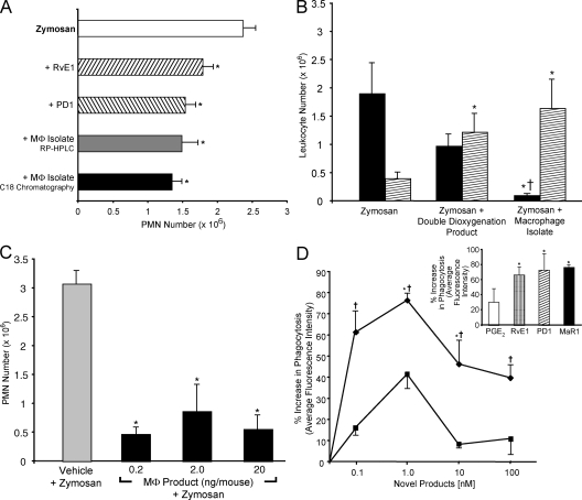
Figure 3.
Antiinflammatory and proresolving novel MΦ products. (A) Reduction in PMN in peritonitis. Activity in methyl formate fractions from C18 extraction of isolated MΦs and 20 ng/mouse of MΦ products isolated with RP-HPLC, 20 ng/mouse PD1, or 20 ng/mouse RvE1. Results are expressed as exudate PMN means ± SEM (n = 3; *, P < 0.05 compared with zymosan plus vehicle). (B) Differential PMN versus monocyte actions. Mice were injected with 0.1 ng/mouse of the double dioxygenation product, 0.1 ng/mouse of MΦ isolate, or vehicle alone (as in A), followed by i.p injection of 1 mg zymosan to evoke peritonitis. After 2 h, leukocytes were enumerated (black bar, PMNs; hatched bar, mononuclear cells). Results are means ± SEM (n = 3; *, P < 0.05 compared with zymosan plus vehicle; †, P < 0.05 for double dioxygenation vs. MΦ isolate). (C) Reduction in peritonitis showing dose response. MΦ product isolated after HPLC isolation was injected i.v. ∼2 min before i.p. zymosan. Results are means ± SEM (n = 3; *, P < 0.05 compared with zymosan plus vehicle). (D) MaR1 enhances phagocytosis. MΦs (24-well plate, 105 cells/well) were exposed to the indicated concentrations for 15 min followed by FITC-labeled zymosan for 30 min at 37°C. Results are means ± SEM expressed as the percent increase above vehicle (n = 3; *, P < 0.05 compared with vehicle; †, P < 0.05 for double dioxygenation vs. MaR1). The closed diamond represents MaR1, and the closed square represents the double dioxygenation product 7S,14S-diHDHA. (inset) Comparison of MaR1 with other mediators (1 nM).

