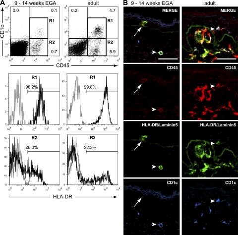
Figure 4.
Not all HLA-DR+ epidermal leukocytes express CD1c at 9 wk EGA. (A) Flow cytometric analysis revealed the presence of CD45+CD1c+ cells in embryonic skin. Histograms display HLA-DR staining (black line) of CD45+CD1c+ (R1) and CD45+CD1c− (R2). Gray line, isotype control. Shown are representative dot plots of three to five experiments per group. (B) Immunofluorescence triple labeling identified HLA-DR+CD1c− (arrows) and HLA-DR+CD1c+ (not depicted) leukocytes in embryonic epidermis. Arrowheads denote CD45+HLA-DR+CD1c+ cells. Data are representative of at least three experiments per group. Bars, 50 μm.

