Figure 8.
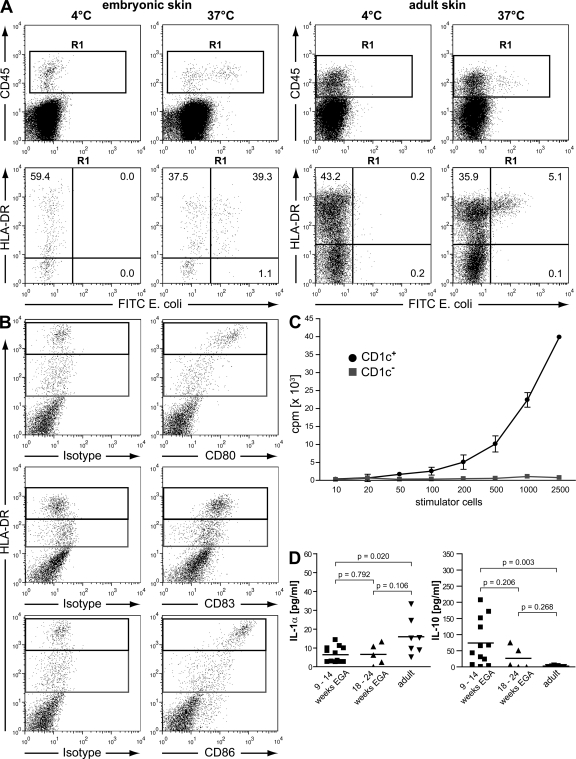
Embryonic DC precursors ingest E. coli, up-regulate costimulatory molecules during culture, and stimulate allogeneic T cells. (A) Phagocytic capacity of leukocytes from embryonic (9–14 wk EGA) and adult skin was assessed by the uptake of FITC-labeled E. coli (107 bacteria/2 × 106 cells). Flow cytometric analysis revealed exclusive uptake of bacteria in CD45+HLA-DR+ cells (R1). Data are representative of three experiments in embryonic skin and of three experiments in adult skin. (B) Single cells from embryonic skin were cultured for 48 h and expression of CD80, CD86 (donor 1), and CD83 (donor 2) on HLA-DR+ cells was analyzed (gated on CD45+ cells). Shown are two representative experiments out of three to five. (C) Allogeneic T cells (5 × 10⁴/well) were cocultured with graded numbers of CD1c+ and CD1c− stimulator cells from embryonic skin. T cell proliferation was determined after 5 d by measuring the incorporation of [3H]thymidine. Data are displayed as the mean ± SD of triplicate cultures, and one representative of three independent experiments is shown. (D) Single cell suspensions of embryonic, fetal, and adult skin were cultured for 48 h, and IL-1α and IL-10 levels in supernatants were analyzed by Luminex technology. Bars represent the mean of investigated groups. 9-14 wk EGA: IL-1α and IL-10, n = 12 each. 18–24 wk EGA: IL-1α and IL-10, n = 5 each. Adult: IL-1α and IL-10, n = 7 each.

