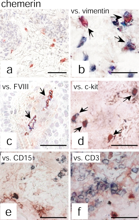
Figure 2.
Chemerin expression colocalizes with vimentin+, FVIII+, and c-kit+ cells. Chemerin expression was evaluated by double immunohistochemistry in psoriatic plaque lesions. Anti-chemerin mAb (red) immunoreactivity was detected in dermal cells having fibroblast-like morphology (a). Double-staining analysis revealed that chemerin colocalizes with vimentin (b), FVIII (c), and c-kit (d) but not with CD15 (e) or CD3 (f) staining (all blue). The figure shows the staining of one biopsy that is representative of eight different patients evaluated. Arrows indicate cells that are double positive for chemerin and for vimentin (b), FVIII (c), or c-kit (d). Bars, 20 μm.

