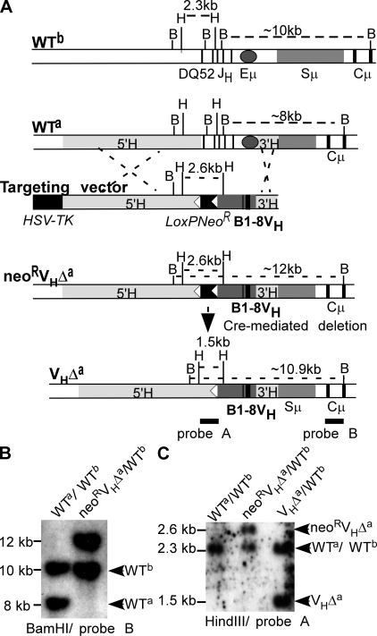
Figure 1.
Eμ deletion and VH gene insertion in an Igha locus. (A) Targeting strategy. WTb, WT Ighb locus. Thick vertical bars show exons for DQ52, JH1-4, and the first two exons of Cμ. Sμ, μ switch region; shaded oval, Eμ; B, BamHI; H, HindIII; WTa, unmodified Igha locus. Shaded boxes (5′H and 3′H) show regions of homology with targeting vector. Targeting vector: LoxPNeoR, loxP-flanked neomycin resistance gene (white arrowheads indicate loxP sites), B1-8VH promoter region (shaded), and coding sequences (thick vertical lines). neoRVHΔa: Igha allele after homologous recombination. VHΔa, modified Igha allele after neoR gene deletion. (B) Southern blot of liver DNA from WT (WTa/WTb) and mouse heterozygous for modified Igha allele before neoR deletion (neoRVHΔa/WTb). (C) Southern blot of liver DNA from WTa/WTb, neoR VHΔa/WTb, and heterozygous mutant mice after neoR deletion (VHΔa/WTb).

