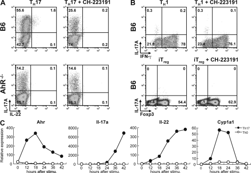
Figure 1.
Increased Th17 polarization caused by endogenous AhR ligands. (A) Intracellular staining for IL-17 versus IL-22 of CD4 T cells from control B6 (top) or AhR-deficient (bottom) cells polarized under Th17 conditions in IMDM for 5 d in the absence (left) or presence (right) of AhR antagonist CH-223191. Representative dot plots of four independent experiments are shown. (B) Intracellular staining for IL-17A versus IFN-γ or IL-17A versus Foxp3 of CD4 T cells from B6 mice cultured under Th1 conditions (top) or iT reg cell conditions (bottom) in the absence (left) or presence (right) of AhR antagonist CH-223191. Dot plots are representative of three independent experiments. (C) qPCR kinetic analysis of AhR, IL-17, IL-22, and CYP1A1 expression after activation of naive CD4 T cells under Th17 conditions. Expression in CD4 T cells activated under neutral conditions is shown for comparison. The figure shows mRNA levels normalized to Hprt expression and is representative of three independent experiments.

