FIG. 3.
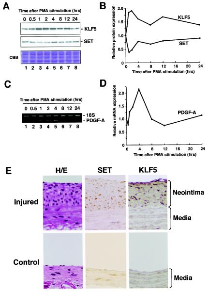
Effects of SET on KLF5 downstream gene expression and pathological states. (A) Induction of KLF5 protein and repression of SET protein after mitogenic stimulation. Cells were starved at the times shown for 24 h, incubated with 100 ng of PMA/ml for the indicated times, and then harvested. Cell lysate was resolved by SDS-PAGE and subjected to Western blotting or Coomassie brilliant blue staining. (B) Quantification of KLF5 and SET protein levels. KLF5 and SET protein levels were normalized by the corresponding Coomassie brilliant blue staining pattern. The relative expression level was shown as the level at 0 h. (C) Induction of PDGF-A chain mRNA expression. Cells were starved at the times shown for 24 h, incubated with 100 ng of PMA/ml for the indicated times, and then harvested. The quantitative reverse transcription-PCR fragmentwas resolved on a 2% agarose gel. (D) Quantification of mRNA expression level for the PDGF-A chain. The expression level of the PDGF-A chain, an endogenous target gene of KLF5, was normalized to that of 18S. (E) KLF5 and SET expression in the pathological neointima. The immunohistochemistry of SET and KLF5 in a balloon injury model of atherosclerosis was examined. The left common carotid artery was denudated by balloon injury, and the neointima was observed 2 weeks after the balloon injury (Injured). The right common carotid artery served as a control (Control). The rat aorta was stained with anti-SET and anti-KLF5 antibodies. Cells in the neointima were clearly positive for SET and KLF5. All experiments were done at least twice with consistent findings. H/E, hematoxylin and eosin staining.

