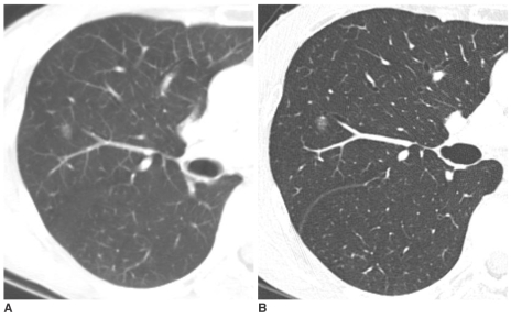
Fig. 1.
Pure nodular ground-glass opacity in the right upper lobe confirmed as atypical adenomatous hyperplasia in a 62-year-old woman.
A. Initial CT shows 8 mm pure nodular ground-glass opacity in the right upper lobe.
B. Thin-section CT after 10 months shows persistent pure nodular ground-glass opacity with the same size. The lesion was pathologically confirmed by right upper lobectomy. She had a history of curative resection of adenocarcinoma in the left upper lobe one year before.

