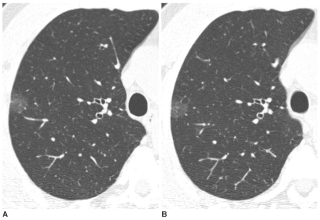
Fig. 2.
Pure nodular ground-glass opacity in the right upper lobe confirmed as atypical adenomatous hyperplasia in a 58-year-old man.
A. Initial thin-section CT shows a 15 mm pure nodular ground-glass opacity in the right upper lobe.
B. On thin-section CT after 2 months, an interval change was not noted. An additional two pure nodular ground-glass opacities were found in the right upper lobe and one in the right middle lobe. All lesions were pathologically confirmed as atypical adenomatous hyperplasia by multifocal wedge resection.

