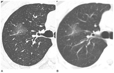
Fig. 4.
Pure nodular ground-glass opacity in the right upper lobe confirmed as focal interstitial fibrosis in a 61-year-old man.
A. Thin-section CT shows 30 mm pure nodular ground-glass opacity in the right upper lobe.
B. On follow-up CT scan after seven months, an interval change was not noted. The lesion was pathologically confirmed by right upper lobectomy. On pathologic slides, focal interstitial fibrosis with exuberant type II pneumocyte proliferation and alveolar macrophage collection were found. The cause of the focalinterstitial fibrosis was not confirmed.

