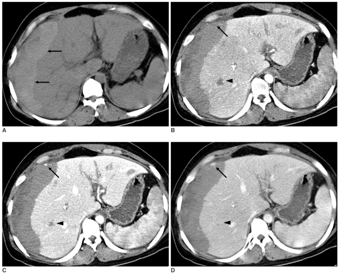
Fig. 3.
The unenhanced CT scan shows massive acute hematoma in the right subcapsular area (A, arrows). The triphasic, contrast-enhanced CT scan shows small enhancing focus (arrow) on the background of hemorrhage, which is isodense to liver parenchyma on the arterial (B), portal (C) and delayed (D) phases. Note a small lesion in the right lobe (arrowhead) that is hypodense on the arterial phase (B), peripherally enhanced on the portal venous phase (C), and it becomes totally isodense or slightly hyperdense on the delayed phase (D).

