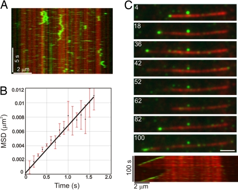
Fig. 2.
CLIP-170 requires EB1 for +TIP activity. (A) Kymograph showing the microtubule binding and diffusive movement of CLIP-170(H2)–GFP at 25 nM. Note the transient binding of CLIP-170(H2)–GFP along the length of the microtubule lattice. (B) Plot of the mean squared displacement (MSD) of CLIP-170(H2)–GFP against time. A linear fit to the data yields a diffusion coefficient, D, of 3.5 ± 0.1 × 10⁻¹¹ cm2 s−1 (MSD = 2Dt). Error bars represent the SEMs of the squared displacement values (n = 38). (C) The montage at the top depicts the specific decoration of growing microtubule plus-ends by 25 nM CLIP-170(H2)–GFP in the presence of 250 nM of unlabeled EB1 protein. The kymograph at the bottom shows the same microtubule over the duration of the observation period. Microtubule depolymerization is accompanied by loss of CLIP-170(H2)2–GFP at the plus-ends and subsequent rescue of microtubule growth restores plus-end tracking of CLIP-170(H2)–GFP. The numbers on each frame represent time (in seconds). (Scale bar, 2 μm.)

