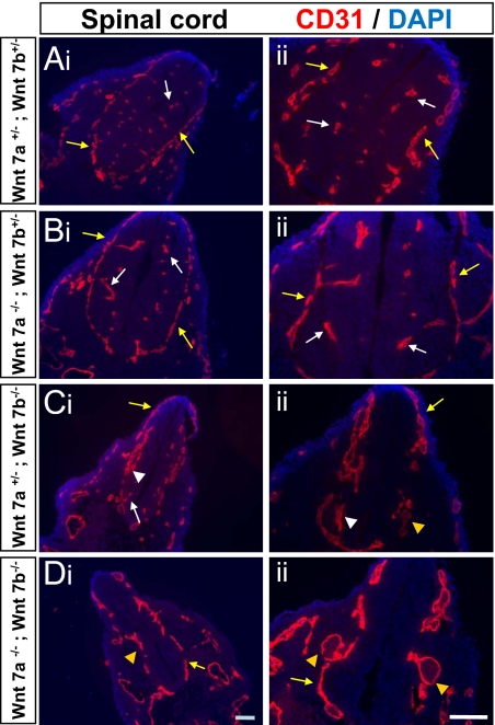
Fig. 4.
Abnormal vasculature in the CNS of Wnt 7 mutants. (A-D) Coronal tissue sections of the E10.5 spinal cord in Wnt7a, Wnt7b double heterozygotes (A: Wnt 7a+/−; Wnt 7b+/−), Wnt 7a mutants (B: Wnt 7a−/− ;Wnt 7b+/−), Wnt 7b mutants (C: Wnt 7a+/− ;Wnt 7b−/−), and Wnt7a, Wnt 7b double mutants (D: Wnt 7a−/− ;Wnt 7b−/−) were stained with the nuclear marker DAPI (blue) and the vascular marker CD31 (red). Normal capillary beds were observed in the wild-type and Wnt7a mutants, whereas vascular malformations and thickened vascular plexus were observed in the Wnt7b mutants, and large vascular plexus dilations were observed in double mutants. Normal capillaries and normal vascular plexus are indicated with white and yellow arrows respectively, whereas vascular malformations and abnormal vascular plexus are indicated with white and yellow arrow heads respectively. (Scale bar, 100 μm.)

