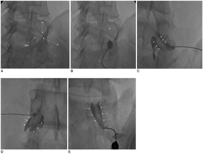
Fig. 1.
Transforaminal epidural steroid injection.
A. Left anterior oblique radiograph for the right L5 transforaminal injection. The triangle is formed by the iliac crest (IC), the inferior margin of the right transverse process (TP), and the right S1 superior articular process (SAP).
B. Left anterior oblique radiograph for needle positioning. The needle tip is inferior and lateral to the right L5 pedicle (p = pedicle).
C. Epidurography shows the outline of the right L5 nerve root sleeve (arrows).
D. Epidurography for the left L4 transforaminal injection. Contrast is seen outlining the left L4 nerve root sleeve (arrows).
E. Epidurography for the right S1 transforaminal injection (arrows).

