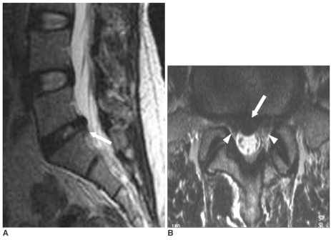
Fig. 3.
MRI of a 66-year-old man. Central disc herniation and grade I nerve root compression with a successful result. Mid-line sagittal (A) and axial (B) T2-weighted MR images show central disc protrusion (arrows). The disc was abutting both S1 nerve roots (arrowheads). Because the distribution of the patient's pain was along the right S1 dermatome and the electrodiagnostic study showed right S1 radiculopathy, we performed right S1 transforaminal ESI. The follow-up assessment seven months after injection showed a pain reduction greater than 50% and a patient satisfaction score of 2.

