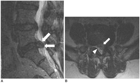
Fig. 6.
MRI of a 46-year-old woman. Grade III nerve root compression with an unsatisfactory result. The paramedian sagittal (A) and axial (B) T2-weighted MR images show a left central extruded disc herniation (arrows). The margin of the herniated disc has moderate signal intensity. The left S1 nerve root is not visible due to the herniated disc, as compared to the right S1 nerve root (arrowhead). Left S1 transforaminal epidural steroid injection was performed, but the symptoms persisted. The patient underwent surgery 1.5 months after injection.

