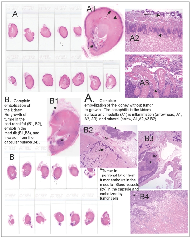
Fig. 6.
Embolized kidneys from Groups 6 and 7 demonstrating the pattern of tumor re-growth.
A. The entire kidney is sectioned, demonstrating a smaller kidney that is completely embolized. No tumor was present in any section (score = 0) on microscopic examination. A2 (magnified x 25) and A3 (magnified x 400) demonstrate that at the capsular surface, the outermost blue/purple is mineralization (arrow), and the inner blue/purple (arrowhead) at the junction with the necrotic parenchyma is inflammation.
B. The entire kidney is sectioned and this includes some adherent peri-renal fat containing blue/purple nodules (*) that are VX2 tumors. In B2 (magnified x 25), the tumor (*) is in the peri-renal fat, and the fat is separated from the renal parenchyma by blood vessels (bv) in the granulating renal capsule. The blue/purple (arrow) below the capsule is mineral on microscopy. B3 (magnified x 25) is a tumor (*) in the medulla of the same section that appears to arrive from a tumor's embolized vessel (bv). B4 (magnified x 100) demonstrated that the tumor (*) invades from the capsular surface into the pink embolized renal parenchyma.

