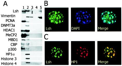
FIG. 4.
Lsh localizes to pericentromeric heterochromatin. (A) Lsh predominantly associates with chromatin. 3T3 cells were extracted with Triton X, and different fractions examined by Western analysis with the indicated antibodies: lane 1, flushed fraction; lane 2, Triton X-resistant fraction after DNase digestion; lane 3, ammonium sulfate wash; lane 4, 2 M NaCl wash; lane 5, solubilized pellet. (B) Lsh colocalizes with DAPI. At 24 h after transfection, GFP-tagged Lsh was examined by fluorescence microscopy. (C) Lsh colocalizes with HP1α. At 24 h after transfection, GFP-tagged Lsh was immunostained for detection of HP1α.

