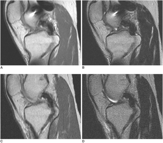
Fig. 1.
Proton density-weighted sagittal (A, C) (TR/TE, 2000/20 msec; echo train length, 4) and T2-weighted sagittal (B, D) (TR/TE, 2000/80 msec; echo train length, 4) images of a 21-year-old man who received posterior cruciate ligament reconstruction using autogenous double-loop hamstring tendon (patient #7). Proton density-weighted image obtained 232 days after posterior cruciate ligament reconstruction shows high band-like peripheral signal intensity (arrowheads) in the proximal and middle segments (A). The high signal intensity has disappeared, and the graft shows homogeneous low signal intensity on proton density-weighted image obtained 472 days after posterior cruciat ligament reconstruction (C). The graft shows homogenous low signal intensity on T2-weighted images (B, D).

