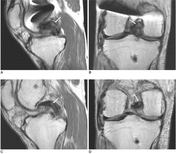
Fig. 2.
Proton density-weighted sagittal (A, C) and coronal (B, D) images (TR/TE, 2000/20 msec; echo train length, 4) of a 35-year-old man who received posterior cruciate ligament reconstruction using autogenous double-loop hamstring tendon graft (patient #6). MR images obtained 265 days after posterior cruciate ligament reconstruction (A, B) show high band-like intrasubstance signal intensity (arrows) in the middle segment. The high signal intensity (arrows) persists on MR images obtained 407 days after posterior cruciate ligament reconstruction (C, D). The graft thickness decreases from 15 mm (A) to 12 mm (C).

