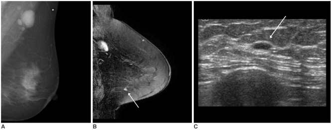
Fig. 3.
A 53-year-old woman with palpable left axillary lymph node metastasis. The mammogram (A) and US showed no abnormal findings in the breast. The contrast-enhanced MR image (B) showed 5 mm nodular enhancement without a washout pattern (arrow) in the left lower outer breast. On the MR-guided second-look US examination (C), a well-defined flat nodular lesion (arrow) was identified, and invasive ductal carcinoma was diagnosed by US-guided localization and excision.

