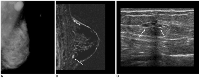
Fig. 4.
A 42-year-old woman with palpable left axillary lymph node metastasis. The mammogram (A) showed extremely dense breast parenchyma without abnormal findings in the breast. The initial screening US was normal. The standard subtraction image (B) of the contrast-enhanced breast MRI showed an enhancing nodule, about 7 mm in size, in the left lower breast (arrow) with a washout pattern. The MR-guided second look US examination localized a few benign cysts in that area (arrows) (C). US-guided fine needle aspiration revealed malignant cells.

