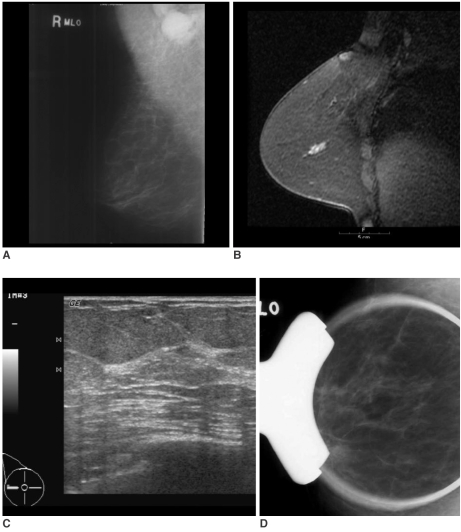
Fig. 5.
A 57-year-old woman with palpable right axillary lymph node metastasis. The mammogram (A) and US showed negative findings in the breast. The standard subtraction image of the contrast enhanced-MRI (B) showed a 1.8-cm sized area of linear enhancement with an early washout pattern, suggesting malignancy in the outer portion of the right breast. MR-guided second-look US (C) and spot-compression with magnification mammography (D), which targeted the areas of MR-detected lesion through the use of a vitamin E capsule attached to the surface of the right breast overlying the MR-detected lesion, could not find the corresponding lesion. After modified radical mastectomy, ductal carcinoma in situ (0.9 cm in extent) was found in the right outer breast.

