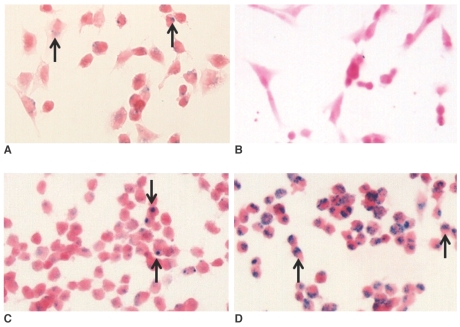
Fig. 1.
Photomicrographs of the hNSCs treated for 24 hr with ferumoxides (A), MION-47 (B), CLIO-NH2 (C) or tat-CLIO (D) at 25 µg/ml. The intracellular uptake of iron oxide nanoparticles (arrows) is seen in cells exposed to ferumoxides (A), CLIO-NH2 (C) or tat-CLIO (D). However, no intracellular uptake of iron oxide was found for the cells incubated with MION-47 (B). (Prussian blue stain, objective magnification: × 40)

