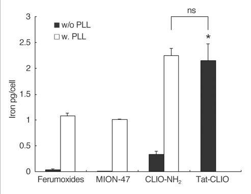
Fig. 4.
Quantitative iron determination by an atomic absorption spectrophotometer. The graph shows the highest iron incorporation in the tat-CLIO labeled cells (2.15 ± 0.3 pg/cell) and the lowest in the MION-47 labeled cells (0.005 pg/cell) in absence of PLL (black bars). With PLL (white bars), the iron content in the ferumoxides labeled, MION-47 labeled or CLIO-NH2 labeled cells increased to 1.08 ± 0.07 pg/cell, 1.01 ± 0.02 pg/cell and 2.24 ± 0.17 pg/cell, respectively, which are 27 fold, 202 fold and 7-fold greater uptakes, respectively, compared with the cells without using PLL. The cells labeled with CLIO-NH2 in the presence of PLL show a similar intracellular iron content as the tat-CLIO-labeled cells (p > 0.1).
Note.-w/o = without, w = with, ns = statistically non-significant

