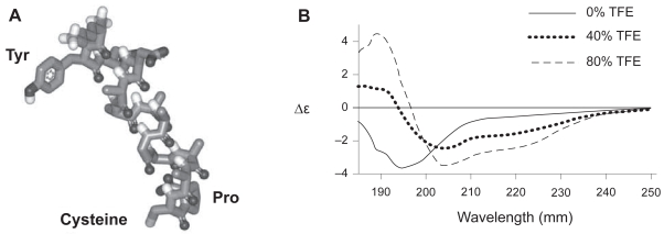
Figure 1.
(A) Image of the predicted structure of the switch tag peptide (ST) in its helical conformation. (B) Far UV CD spectra of ST. A 0.2 mg/ml solution of peptide in 50 mM NaH2PO4 buffer, pH 7.0 was used throughout. Increasing the quantity of the hydrophobic solvent TFE promoted the transition of peptide secondary structure from random coil to α-helix.
Abbreviation: TFE, tri-fluoro ethanol.

