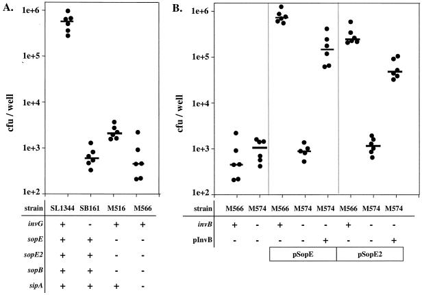
FIG. 11.
Role of invB in SopE/SopE2-mediated host cell invasion. (A) SL1344 (wild type), SB161 (ΔinvG), M516 (ΔsopB sopE sopE2), and M566 (ΔsopB ΔsopE ΔsopE2 ΔsipA) were grown under SPI-1-inducing conditions and used to infect COS-7 tissue culture cells (MOI, 15 bacteria/cell) for 50 min, and the number of internalized bacteria was measured by the gentamicin protection assay (see Materials and Methods). The numbers of CFU per well from six independent experiments are shown. The bars indicate medians. (B) M566 (ΔsopB ΔsopE ΔsopE2 ΔsipA) and M574 (ΔsopB ΔsopE ΔsopE2 ΔsipA invB::aphT) carrying one or more of plasmids pSB1130, pM149, and pM249 were grown under SPI-1-inducing conditions (see Materials and Methods). COS-7 cells were infected as described above. The numbers of intracellular bacteria were determined in at least five independent experiments. The bars indicate medians.

