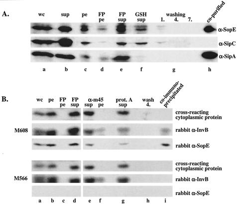
FIG. 3.
(A) Western blot analysis of a GST-InvB affinity purification assay. GST-InvB (pM672) was expressed in wild-type Salmonella serovar Typhimurium SL1344 (see Materials and Methods). The bacterial lysate (25 ml) was incubated with 200 μl of GSH-Sepharose beads. Samples from the purification steps were analyzed by Western blotting by using anti-SopE (α-SopE), anti-SipA (α-SipA), and anti-SipC (α-SipC) antisera. Lane a, 100 μl of a whole culture before harvesting of the cells (wc); lane b, proteins recovered from 250 μl of culture supernatant after pelleting of the cells (sup); lane c, 25 μl of a bacterial pellet resuspended in 25 ml of buffer B; lane d, 50 μl of pelleted cell debris resuspended in 25 ml of buffer B after lysis with a French pressure cell (FP pe); lane e, 50 μl of cleared French pressure cell lysate (total volume, 25 ml) (FP sup); lane f, 50 μl of cleared cell lysate after binding of GST-InvB and its associated proteins to 200 μl of GSH-Sepharose beads (GSH sup); lanes g, 100 μl of washing solution after the first, fourth, and seventh washes of the GSH-Sepharose beads with buffer B (washing 1, 4, and 7, respectively); lane h, 10 μl of GSH-Sepharose beads. (B) Western blot analysis of a coimmunoprecipitation experiment performed with the sopEM45-expressing strain M608 and an anti-M45 antibody (see Materials and Methods). The bacterial lysate (5 ml) was incubated with 10 μl of monoclonal mouse anti-M45 antibody and 10 μl of protein A-Sepharose beads. Samples from the precipitation procedure were analyzed by Western blotting by using polyclonal rabbit anti-InvB (rabbit α-InvB) and anti-SopE (rabbit α-SopE) antisera. Lane a, 100 μl of a whole culture before harvesting of the cells (wc); lane b, 100 μl of bacterial pellet resuspended in 20 ml of buffer B (pe); lane c, 100 μl of pelleted cell debris resuspended in 20 ml of buffer B after lysis with a French pressure cell (FP pe); lane d, 100 μl of cleared French pressure cell lysate (FP sup); lane e, 100 μl of cleared cell lysate after incubation with anti-M45 antibody and removal of nonspecific aggregates by centrifugation (α-m45 sup); lane f, 100 μl of pelleted nonspecific aggregates resuspended in buffer B (α-m45 pe); lane g, 100 μl of cleared cell lysate after incubation with 10 μl of protein A-Sepharose beads (prot. A sup); lane h, 100 μl of washing solution after the fourth wash of the protein A-Sepharose beads with buffer B (wash 4); lane i, 10 μl of protein A-Sepharose beads.

