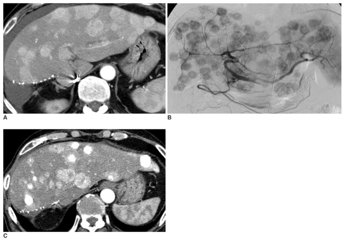
Fig. 2.
A.Contrast-enhanced delayed-phase axial CT image of a transplanted liver six months after living donor liver transplantation, with the left lobe showing multiple recurrent hepatocellular carcinomas in the entire transplanted liver; however, blood flow is preserved in the portal vein.
B.Arteriogram from the celiac trunk (late arterial phase) shows multiple hypervascular tumors in the entire transplanted liver.
C.Contrast-enhanced arterial-phase axial CT image of the transplanted liver one month after transcatheter arterial chemoembolization shows progression of the recurrent hepatocellular carcinomas in both size and number. Some nodules show partial accumulation of iodized oil. We interpreted this patient as having progressive disease.

