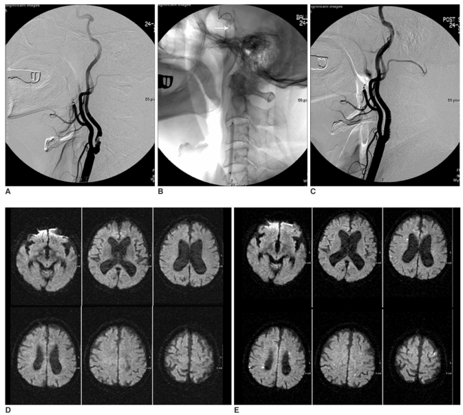
Fig. 1.
A 72-year-old man who underwent protected carotid artery stenting with a balloon device.
A. Pre-stenting angiogram shows severe stenosis (86.3%) at the left internal carotid artery.
B. A balloon device is deployed in the distal carotid artery (arrow).
C. After carotid artery stenting, the lumen of the left internal carotid artery is successfully dilated.
D. No ischemic lesion is shown in bilateral cerebral hemispheres on the pre-stenting diffusion weighted MR imaging.
E. Multiple small new hyperintensities are shown on the post-stenting diffusion-weighted MR imaging. Note the new hyperintnesities are distributed in not only the ipsilateral but also the contralateral cerebral hemisphere. However, no symptomatic neurological complications occurred after carotid artery stenting.

