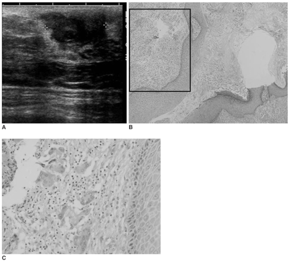
Fig. 1.
A 50-year-old woman with a palpable left subareolar mass.
A. Sonography shows a heterogeneoulsy hypoechoic mass with an indistinct margin.
B. Photomicrograph shows that the lesion is lined by epidermal-type epithelium and it is filled with keratinous material, which is pathognomonic for an epidermal inclusion cyst. (Hematoxylin & Eosin staining × 40)
C. Photomicrograph of adjacent tissue (box in B) reveals inflammatory infiltrate cells or a foreign body reaction. (Hematoxylin & Eosin staining × 100)

