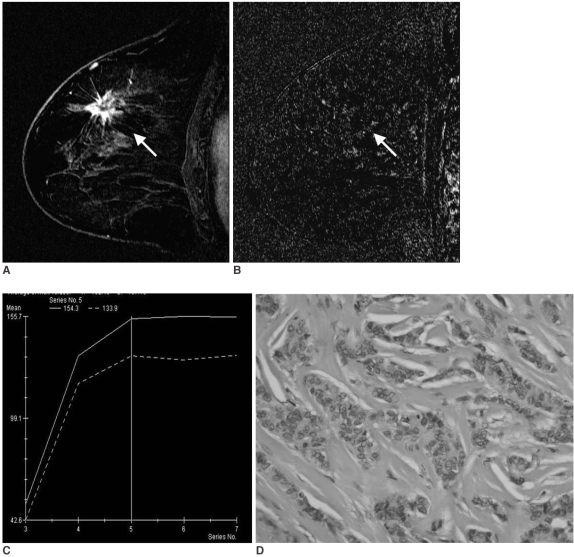
Fig. 1.
A 56-year-old woman with invasive ductal carcinoma not otherwise specified of histological grade 1, ER (+), PR (+), Ki-67 (-).
A. Sagittal standard subtracted fast lowangle shot MR image shows an irregular mass with a spiculated margin (arrow).
B. Sagittal reverse subtracted fast lowangle shot MR image shows nonwashout kinetics (arrow).
C. Time-signal intensity curve shows plateau late enhancement.
D. Photomicrograph shows high proportional tubule formation, low nuclear pleomorphism, and a low mitotic count (Hematoxylin & Eosin staining, ×400).

