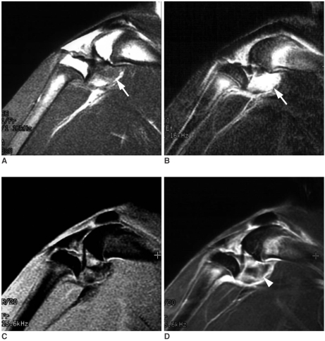
Fig. 3.
MR images of carrageenin-induced arthritis of the rabbit.
Sagittal scan of T1-weighted image (A), fat-suppressed, fast spin-echo T2-weighted image (B), three-dimensional fat-suppressed, fast spoiled gradient-echo (3D FS-FSPGR) image before contrast enhancement (C), and 3D FS-FSPGR image after contrast enhancement (D) show joint effusion with posterior joint capsular distension (arrow in A and B) and thick contrast enhancement at the posterior margin of the joint space on the 3D FS-FSPGR image (arrowhead in D, grade 2).

