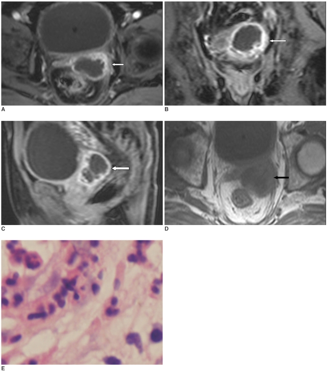
Fig. 1.
Seminal vesicle cyst infection in 78-year-old man.
A-D. Pelvic MRI demonstrates that cystic wall of lesion in left seminal vesicle is irregularly thick, and it is enhanced markedly and inhomogenous on axial, coronal and sagittal T1-weighted enhanced images (A-C) (white arrows), as compared with depiction on axial T1-weighted image (D) (black arrow).
E. Photomicrography of histological specimen shows inflammatory corpuscles composed of lymphocytes, neutrophilic leukocytes and plasmocytes (×40).

