Fig. 1.
Congenital coronary sinus anomaly in 60-year-old woman with Ebstein's anomaly.
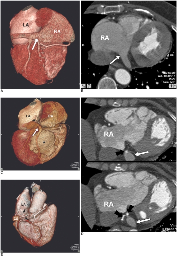
A, B. Dorsal view of reconstructed volume-rendered image (A) and maximum-intensity-projection image (B) reveal normal coronary sinus (arrow) drain into right atrium (RA). RA = right atrium; LA = left atrium
C. Dorsal view of reconstructed volume-rendered image reveals abnormal engorged coronary sinus (long white arrow) without gross communication with right atrium (RA). There was small tortuous vascular channel (short black arrow) drain into left atrium (LA). Atrialization portion of right ventricle also could be identified (black star).
D. Series maximum-intensity-projection images confirmed that there was no communication between abnormally engorged coronary sinus (long white arrows) and right atrium (RA). In addition, there was small tortuous vascular channel with high contrast density (black arrows) located between engorged coronary sinus and right atrium (RA). Small foci of high contrast density within coronary sinus also had been found (black arrows).
E. Post-processing oblique-dorsal view of reconstructed volume-rendered image with total removal of right atrium revealed that this small tortuous vascular channel (black arrows) connected coronary sinus (CS) and left atrium (LA).
RA = right atrium; LA = left atrium; CS = coronary sinus

