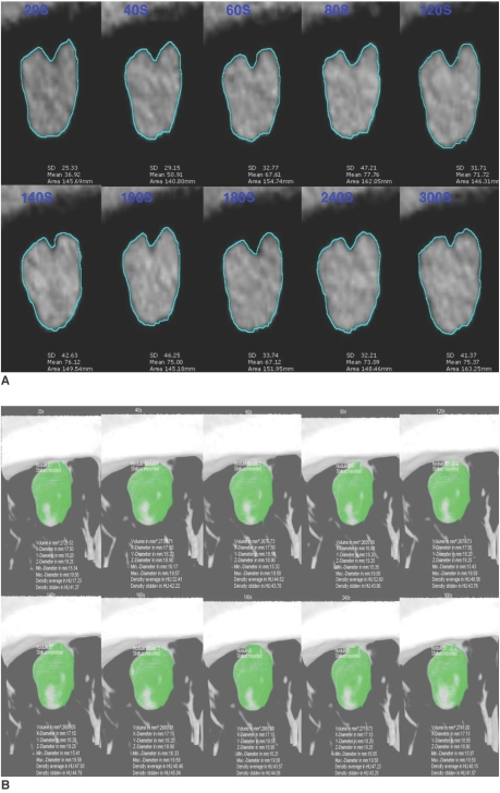
Fig. 1.
Methods of enhancement pattern analysis using CAD system.
A. Before intravenously injecting contrast medium, series of images was obtained throughout entire nodule along z-axis, and additional ten sets (20s, 40s, 60s, 80s, 120s, 140s, 160s, 180s, 240s and 300s) of images were obtained at 20-second intervals over 5-minute period after injecting contrast medium. Region of interest covered approximately full diameter of nodule and single radiologist directly drew region of interest.
B. Solitary pulmonary nodule was region of interest drawn by expert technician with using CAD system.

