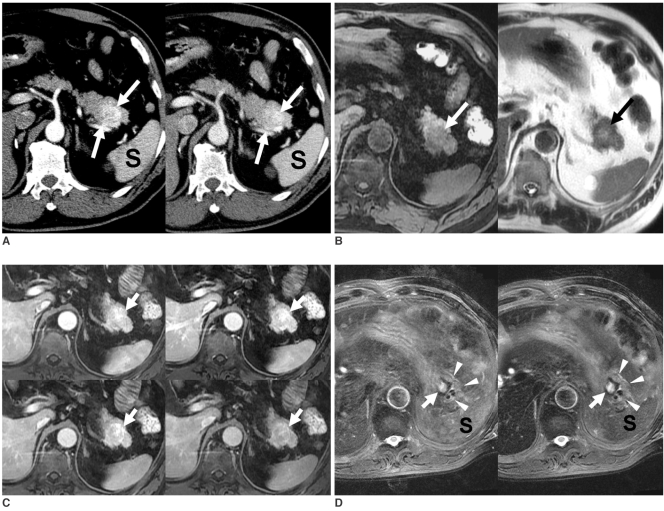
Fig. 10.
Solitary pancreatic metastasis from renal cell carcinoma that occurred six years after right nephrectomy.
A. On arterial phase CT scan (left), lesion (arrows) enhanced more strongly than did spleen (S). Delayed phase CT scan (right) reveals slightly higher attenuation of lesion (arrows) compared to that of pancreas.
B. Lesion (arrows) is hypointense and heterogeneous hyperintense compared to pancreas on T1- (left) and T2-weighted (right) MR images, respectively.
C. On serial dynamic Gd-enhanced MR images obtained at similar phases to those in Figure 9, metastasis (arrows) demonstrates enhancement similar to that described on CT.
D. Precontrast (left) and SPIO-enhanced (right) fat-saturated T2-weighted images do not show any signal drop of lesion (arrows) in pancreas (arrowheads). Note signal drop of spleen (S) on SPIO-enhanced image (right) when compared to precontrast image (left).

