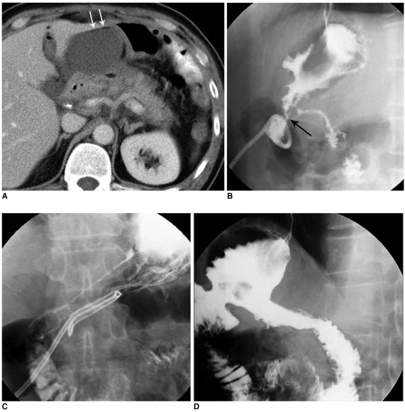
Fig. 3.
High-output fistula after Billroth I operation due to stomach cancer.
A. Abdomen CT image shows abnormal loculated fluid collection with scanty air-bubbles (arrows) adjacent to gastroduodenal anastomotic site.
B. Fistulogram after insertion of drainage tube shows fistula tract (arrow) from intra-abdominal abscess pocket to remnant of stomach via anastomosis site and stenosis at anastomosis site.
C. Upper GI series obtained immediately after placement of covered Nitinol stent at gastroduodenal anastomosis site shows fully expanded stent with good passage of contrast media without visible fistula tract.
D. Follow-up upper GI series taken three months after stent placement shows properly located stent with excellent patency.

