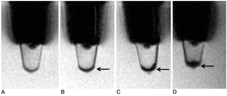
Fig. 2.
In vitro T2*-weighted images of neural stem cells.
A-D. 250 neural stem cells in 2% gelatin tube (A), 5 × 10³ neural stem cells (B), 5 × 10⁴ neural stem cells (C), 2 × 10⁵ neural stem cells (D). In B, C, and D, T2*-weighted images (TR/TE = 231/10 msec) show linear, hypointense cluster in bottom of tubes (arrows), representing SPIO-labeled neural stem cells.

